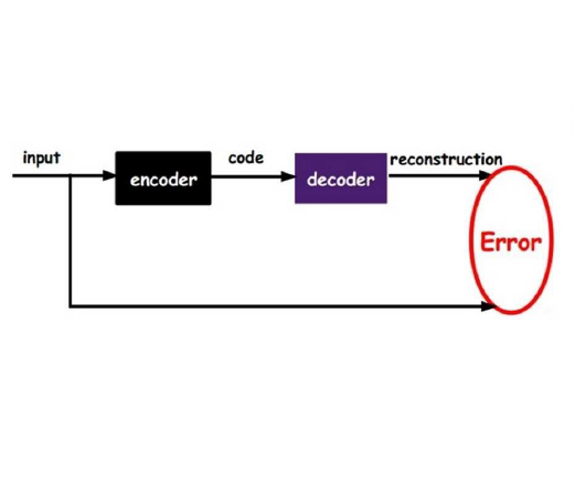
In this work, we propose a two-stage autoencoder based compressor-decompressor framework for compressing malaria RBC cell image patches. We know that the medical images used for disease diagnosis are around multiple gigabytes size, which is quite huge. The proposed residual-based dual autoencoder network is trained to extract the unique features which are then used to reconstruct the original image through the decompressor module. The two latent space representations (first for the original image and second for the residual image) are used to rebuild the final original image. Color-SSIM has been exclusively used to check the quality of the chrominance part of the cell images after decompression. The empirical results indicate that the proposed work outperformed other neural network related compression technique for medical images by approximately 35%, 10% and 5% in PSNR, Color SSIM and MS-SSIM respectively. The algorithm exhibits a significant improvement in bit savings of 76%, 78%, 75% & 74% over JPEG-LS, JP2K-LM, CALIC and recent neural network approach respectively, making it a good compression-decompression technique.
翻译:在这项工作中,我们提出了一个基于压缩疟疾 RBC 细胞图像的基于压缩的压缩器压缩器-减压器的两阶段自动解压缩器框架。 我们知道用于疾病诊断的医疗图像是围绕多个千兆字节大小的, 规模相当大。 拟议的残留的双自动解压缩器网络经过培训, 以提取独有的特性, 这些特性随后通过减压器模块用于重建原始图像。 两种潜在空间显示( 最初图像的1个, 剩余图像的2个) 用于重建最后的原始图像。 色- SSIM 专门用于检查细胞图像的色度部分质量。 彩色- SSIM 已被专门用于检查降压后细胞图像的色度部分。 实证结果表明, 拟议的工作优于其他与神经网络相关的医疗图像压缩技术, PSNRR、 Colory SSIM 和 MS-SSIM 。 算法显示比JEG- LS、 JP2K- LM、 CALIC 和最近的神经网络方法分别提高了74%和74%, 使它成为良好的压缩技术。


