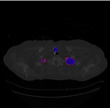
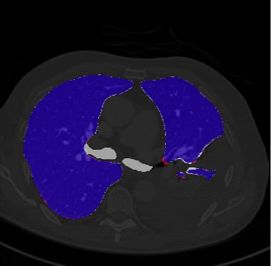
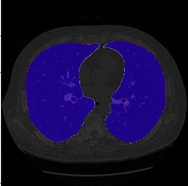
Segmentation of lung tissue in computed tomography (CT) images is a precursor to most pulmonary image analysis applications. Semantic segmentation methods using deep learning have exhibited top-tier performance in recent years. This paper presents a fully automatic method for identifying the lungs in three-dimensional (3D) pulmonary CT images, which we call it Lung-Net. We conjectured that a significant deeper network with inceptionV3 units can achieve a better feature representation of lung CT images without increasing the model complexity in terms of the number of trainable parameters. The method has three main advantages. First, a U-Net architecture with InceptionV3 blocks is developed to resolve the problem of performance degradation and parameter overload. Then, using information from consecutive slices, a new data structure is created to increase generalization potential, allowing more discriminating features to be extracted by making data representation as efficient as possible. Finally, the robustness of the proposed segmentation framework was quantitatively assessed using one public database to train and test the model (LUNA16) and two public databases (ISBI VESSEL12 challenge and CRPF dataset) only for testing the model; each database consists of 700, 23, and 40 CT images, respectively, that were acquired with a different scanner and protocol. Based on the experimental results, the proposed method achieved competitive results over the existing techniques with Dice coefficient of 99.7, 99.1, and 98.8 for LUNA16, VESSEL12, and CRPF datasets, respectively. For segmenting lung tissue in CT images, the proposed model is efficient in terms of time and parameters and outperforms other state-of-the-art methods. Additionally, this model is publicly accessible via a graphical user interface.
翻译:计算断层成像(CT) 图像中的肺组织分解是大多数肺部图像分析应用的先导。 使用深层学习的静态分解方法近年来表现出顶级性能。 本文展示了一种完全自动的三维( 3D) 肺部CT 图像中肺部识别方法, 我们称之为 Lung- Net 。 我们推测, 一个具有初始V3 单位的更深网络可以更好地体现肺部CT 图像的特征, 而不会增加可训练参数数量的模型复杂性。 该方法有三个主要优势。 首先, 开发了一个带有“ 感官V3” 图像块的 U- 网络结构, 以解决性能退化和参数过载的问题。 然后, 利用连续切三维( 3D) 肺部CT) 肺部CT 图像中的信息, 创建了一个新的数据结构, 以便通过尽可能高效的数据表达方式, 来提取更多的偏差。 最后, 利用一个公共数据库, 培训和测试该模型( LUNA16) 和两个公共数据库( ISBI VER- CREL 12 值 和 CRPFS 4 4 4 4 4 4 ) 系统 系统 系统 系统 等 系统 系统 的系统,, 系统 系统 系统 系统 系统 系统 系统 系统 系统 系统 系统 系统 系统 系统 系统 系统 系统 系统 系统 系统 系统 系统 系统 系统 系统 系统 系统 系统 系统 系统 系统 系统 系统 系统 系统 系统 系统 系统 系统 系统 系统 系统 系统 系统 系统 系统 系统 系统 系统 系统 系统 系统 系统 系统 系统 系统 系统 系统 系统 系统 系统 系统 系统 系统 系统 系统 系统 系统 系统 系统 系统 系统 系统 系统 系统 系统 系统 系统 系统 系统 系统 系统 系统 系统 系统 系统 系统 系统 系统 系统 系统 系统 系统 系统 系统 系统 系统 系统 系统 系统 系统 系统 系统 系统 系统 系统 系统 系统 系统 系统 系统 系统


