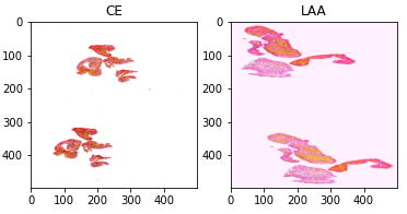
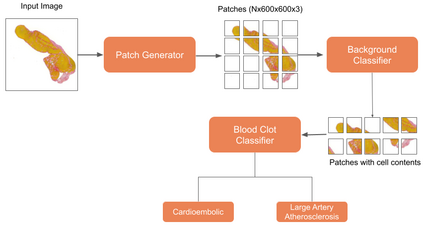
An innovative two-stage methodology for categorizing blood clot origins is presented in this paper, which is important for the diagnosis and treatment of ischemic stroke. First, a background classifier based on MobileNetV3 segments big whole-slide digital pathology images into numerous tiles to detect the presence of cellular material. After that, different pre-trained image classification algorithms are fine-tuned to determine the origin of blood clots. Due to complex blood flow dynamics and limitations in conventional imaging methods such as computed tomography (CT), magnetic resonance imaging (MRI), and ultrasound, identifying the sources of blood clots is a challenging task. Although these techniques are useful for identifying blood clots, they are not very good at determining how they originated. To address these challenges, our method makes use of robust computer vision models that have been refined using information from whole-slide digital pathology images. Out of all the models tested, the PoolFormer \cite{yu2022metaformer} performs better than the others, with 93.4\% accuracy, 93.4\% precision, 93.4\% recall, and 93.4\% F1-score. Moreover, it achieves the good weighted multi-class logarithmic loss (WMCLL) of 0.4361, which emphasizes how effective it is in this particular application. These encouraging findings suggest that our approach can successfully identify the origin of blood clots in a variety of vascular locations, potentially advancing ischemic stroke diagnosis and treatment approaches.
翻译:暂无翻译


