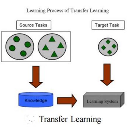
Gliomas are the most common malignant brain tumors that are treated with chemoradiotherapy and surgery. Magnetic Resonance Imaging (MRI) is used by radiotherapists to manually segment brain lesions and to observe their development throughout the therapy. The manual image segmentation process is time-consuming and results tend to vary among different human raters. Therefore, there is a substantial demand for automatic image segmentation algorithms that produce a reliable and accurate segmentation of various brain tissue types. Recent advances in deep learning have led to convolutional neural network architectures that excel at various visual recognition tasks. They have been successfully applied to the medical context including medical image segmentation. In particular, fully convolutional networks (FCNs) such as the U-Net produce state-of-the-art results in the automatic segmentation of brain tumors. MRI brain scans are volumetric and exist in various co-registered modalities that serve as input channels for these FCN architectures. Training algorithms for brain tumor segmentation on this complex input requires large amounts of computational resources and is prone to overfitting. In this work, we construct FCNs with pretrained convolutional encoders. We show that we can stabilize the training process this way and achieve an improvement with respect to dice scores and Hausdorff distances. We also test our method on a privately obtained clinical dataset.
翻译:磁共振成像(MRI)被放射治疗师用于人工切分脑损伤并观察其在整个治疗过程中的发育。人工成像分解过程耗时,结果因人类比例不同而异。因此,对自动成像分解算法的需求很大,这种算法产生可靠和准确的脑组织类型的分解。最近深层次学习的进展导致神经神经网络结构的演进,这些结构在各种视觉识别任务中十分出色。它们已被成功地应用于医学环境,包括医学图像分解。特别是,完全相联的网络(FCNs),例如U-Net产生最新的脑肿瘤自动分解结果。MRI脑扫描是量的,存在于作为FCN结构投入渠道的各种共同注册模式中。关于这种复杂输入的脑肿瘤分解的培训算法需要大量计算资源,并且容易被过度适应。在这种医学环境中,我们用这种分流化方法来稳定大脑分解过程,我们也可以用这种分解的方法来进行我们进化。


