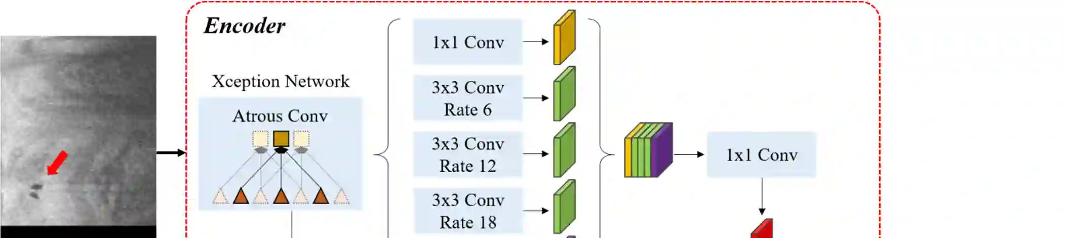
To analyze this characteristic of vulnerability, we developed an automated deep learning method for detecting microvessels in intravascular optical coherence tomography (IVOCT) images. A total of 8,403 IVOCT image frames from 85 lesions and 37 normal segments were analyzed. Manual annotation was done using a dedicated software (OCTOPUS) previously developed by our group. Data augmentation in the polar (r,{\theta}) domain was applied to raw IVOCT images to ensure that microvessels appear at all possible angles. Pre-processing methods included guidewire/shadow detection, lumen segmentation, pixel shifting, and noise reduction. DeepLab v3+ was used to segment microvessel candidates. A bounding box on each candidate was classified as either microvessel or non-microvessel using a shallow convolutional neural network. For better classification, we used data augmentation (i.e., angle rotation) on bounding boxes with a microvessel during network training. Data augmentation and pre-processing steps improved microvessel segmentation performance significantly, yielding a method with Dice of 0.71+/-0.10 and pixel-wise sensitivity/specificity of 87.7+/-6.6%/99.8+/-0.1%. The network for classifying microvessels from candidates performed exceptionally well, with sensitivity of 99.5+/-0.3%, specificity of 98.8+/-1.0%, and accuracy of 99.1+/-0.5%. The classification step eliminated the majority of residual false positives, and the Dice coefficient increased from 0.71 to 0.73. In addition, our method produced 698 image frames with microvessels present, compared to 730 from manual analysis, representing a 4.4% difference. When compared to the manual method, the automated method improved microvessel continuity, implying improved segmentation performance. The method will be useful for research purposes as well as potential future treatment planning.
翻译:为分析这种脆弱性特征,我们开发了一种自动深层学习方法,用于探测血管内光学一致性摄影图像中的微贝。共分析了85个损伤和37个正常部分的8,403个IVOCT图像框。使用本组以前开发的专用软件(OCTOPUS)做了人工批注。在极地(r,theta})域的数据增强应用到原始的 IVOCT图像,以确保微贝出现在所有可能的角度。预处理方法包括导电线/阴影探测、润滑分解、平流变换和减少噪音。DeepLab v3+用于分解微血管候选人。每个候选人的捆绑框被归类为微型船或非微生物,使用浅层神经网络网络网络网络网络网络。为了更好的分类,我们使用数据增强(即角旋转)作为微贝的多数角度。数据增强和预处理前步骤改进了微层分解分解性功能,使0.7.1+0.8的磁性分析方法与0.7+0.8的精确度进行比较。