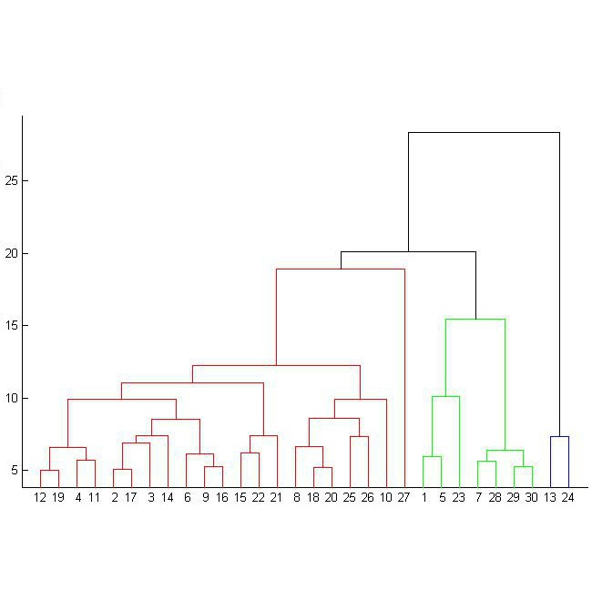
Supervised deep learning has shown state-of-the-art performance for medical image segmentation across different applications, including histopathology and cancer research; however, the manual annotation of such data is extremely laborious. In this work, we explore the use of superpixel approaches to compute a pre-segmentation of HER2 stained images for breast cancer diagnosis that facilitates faster manual annotation and correction in a second step. Four methods are compared: Standard Simple Linear Iterative Clustering (SLIC) as a baseline, a domain adapted SLIC, and superpixels based on feature embeddings of a pretrained ResNet-50 and a denoising autoencoder. To tackle oversegmentation, we propose to hierarchically merge superpixels, based on their content in the respective feature space. When evaluating the approaches on fully manually annotated images, we observe that the autoencoder-based superpixels achieve a 23% increase in boundary F1 score compared to the baseline SLIC superpixels. Furthermore, the boundary F1 score increases by 73% when hierarchical clustering is applied on the adapted SLIC and the autoencoder-based superpixels. These evaluations show encouraging first results for a pre-segmentation for efficient manual refinement without the need for an initial set of annotated training data.
翻译:受监督的深层学习显示,在不同应用中,包括病理学和癌症研究中,医学图像分解的医学图象显示最先进的性能,这包括组织病理学和癌症研究;然而,对这些数据的人工注解极为艰苦。在这项工作中,我们探索使用超像素方法计算乳癌诊断的HER2的染色图像预分层,以便于在第二个步骤中用手动更快的手动注解和校正。比较了四种方法:标准简单线形透析组(SLIC)作为基线,一个区域经调整的 SLIC,以及基于预先训练的ResNet-50和解调自定义自动编码的特征嵌入的超级像素。为了处理超分层分解,我们建议根据各自特性空间的内容,将超像素进行等级合并。在对完全手动的图象进行评价时,我们观察到,基于自动电解的超级像素类比比比SLIC超级像素的边界F1分增加了23%。此外,在应用等级组合前的边界F1分数增加73%,当应用了SLICS-chnicredustrual 之前的高级数据显示一个精化前的高级精化结果。


