Purpose: Lung nodule segmentation, i.e., the algorithmic delineation of the lung nodule surface, is a fundamental component of computational nodule analysis pipelines. We propose a new method for segmentation that is a machine learning based extension of current approaches, using labeled image examples to improve its accuracy. Approach: We introduce an extension of the standard level set image segmentation method where the velocity function is learned from data via machine learning regression methods, rather than a priori designed. Instead, the method employs a set of features to learn a velocity function that guides the level set evolution from initialization. Results: We apply the method to image volumes of lung nodules from CT scans in the publicly available LIDC dataset, obtaining an average intersection over union score of 0.7185($\pm$0.1114), which is competitive with other methods. We analyze segmentation performance by anatomical and appearance-based categories of the nodules, finding that the method performs better for isolated nodules with well-defined margins. We find that the segmentation performance for nodules in more complex surroundings and having more complex CT appearance is improved with the addition of combined global-local features. Conclusions: The level set machine learning segmentation approach proposed herein is competitive with current methods. It provides accurate lung nodule segmentation results in a variety of anatomical contexts.
翻译:目标:肺部结核分离,即肺部结核表面的算法划界,是计算结核分析管道的一个基本组成部分。我们提出一种新的分解方法,这是目前方法的机械学习延伸,使用标签图像示例来提高准确性。方法:我们采用标准水平设置图像分解法的延伸,通过机器学习回归方法而不是先验设计的数据来学习速度函数。相反,该方法使用一套特征来学习一套速度函数,用以指导水平组合从初始化演变。结果:我们采用这种方法,在公开提供的LIDC数据集中,将CT扫描的肺部结核成像量从CT扫描中应用,获得0.7185($\pm$0.1114)的交错平均值,这与其他方法相比具有竞争力。我们用解剖和外观分类方法分析结核的分解性功能,发现该方法对带明确边缘的孤立结核效果更好。我们发现,结核在更复杂的周围和有更复杂的CT外观的分解性表现,随着当前LIDC数据集的增加而得到改进,因此,将得出一个具有准确的磁段级分析结果。
相关内容
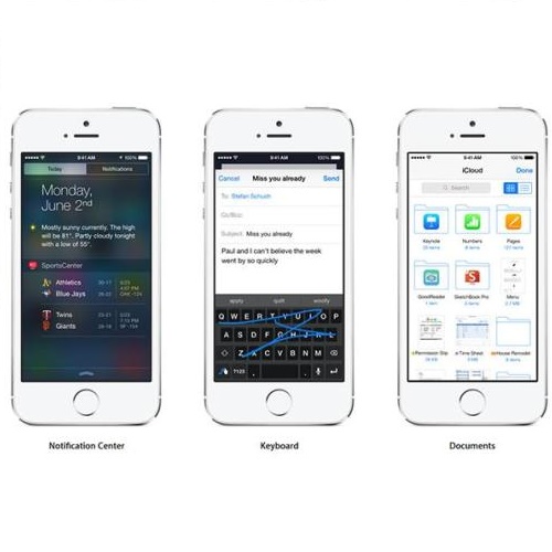
- Today (iOS and OS X): widgets for the Today view of Notification Center
- Share (iOS and OS X): post content to web services or share content with others
- Actions (iOS and OS X): app extensions to view or manipulate inside another app
- Photo Editing (iOS): edit a photo or video in Apple's Photos app with extensions from a third-party apps
- Finder Sync (OS X): remote file storage in the Finder with support for Finder content annotation
- Storage Provider (iOS): an interface between files inside an app and other apps on a user's device
- Custom Keyboard (iOS): system-wide alternative keyboards
Source: iOS 8 Extensions: Apple’s Plan for a Powerful App Ecosystem


