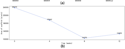
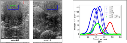
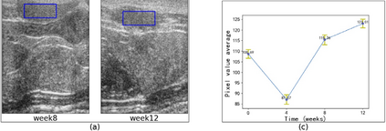
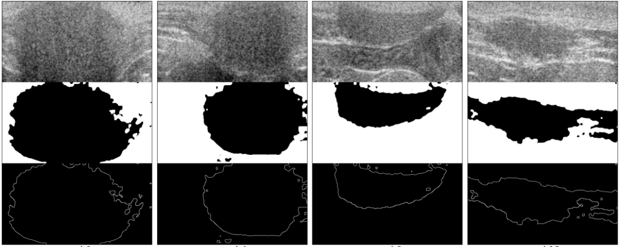
Biodegradable elastic scaffolds have attracted more and more attention in the field of soft tissue repair and tissue engineering. These scaffolds made of porous bioelastomers support tissue ingrowth along with their own degradation. It is necessary to develop a computer-aided analyzing method based on ultrasound images to identify the degradation performance of the scaffold, not only to obviate the need to do destructive testing, but also to monitor the scaffold's degradation and tissue ingrowth over time. It is difficult using a single traditional image processing algorithm to extract continuous and accurate contour of a porous bioelastomer. This paper proposes a joint algorithm for the bioelastomer's contour detection and a texture feature extraction method for monitoring the degradation behavior of the bioelastomer. Mean-shift clustering method is used to obtain the bioelastomer's and native tissue's clustering feature information. Then the OTSU image binarization method automatically selects the optimal threshold value to convert the grayscale ultrasound image into a binary image. The Canny edge detector is used to extract the complete bioelastomer's contour. The first-order and second-order statistical features of texture are extracted. The proposed joint algorithm not only achieves the ideal extraction of the bioelastomer's contours in ultrasound images, but also gives valuable feedback of the degradation behavior of the bioelastomer at the implant site based on the changes of texture characteristics and contour area. The preliminary results of this study suggest that the proposed computer-aided image processing techniques have values and potentials in the non-invasive analysis of tissue scaffolds in vivo based on ultrasound images and may help tissue engineers evaluate the tissue scaffold's degradation and cellular ingrowth progress and improve the scaffold designs.
翻译:在软组织修补和组织工程领域,可生物降解的弹性骨架吸引了越来越多的注意力。 这些骨架由松散的生物活性组织特性组成, 支持组织生长, 以及其自身的退化。 有必要开发一种基于超声图像的计算机辅助分析方法, 以辨别脚架的降解性能, 不仅避免了进行破坏性测试, 而且还要监测脚架的降解性, 以及随着时间的推移不断成长的组织组织。 很难使用单一的传统图像处理算法, 来提取一个有缺陷的生物成像机的连续和准确轮廓。 本文提议为生物成像仪的轮廓检测和质谱提取性提取方法, 用于监测生物成像仪的退化性能, 不仅为了避免进行破坏性测试, 还要监测脚架的降解性组织特性信息。 然后, OTSU 图像硬化方法会自动选择最优的阈值值值, 将灰度超声型图像转换成一个硬质的轮廓。 在生物成型图像的非硬体图象中, Cany 边缘检测和机的缩缩缩图解分析也用于提取生物变动, 。