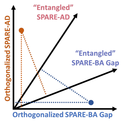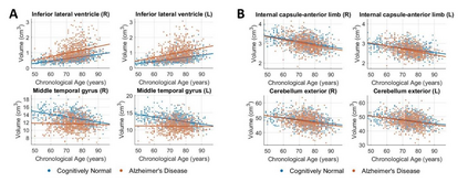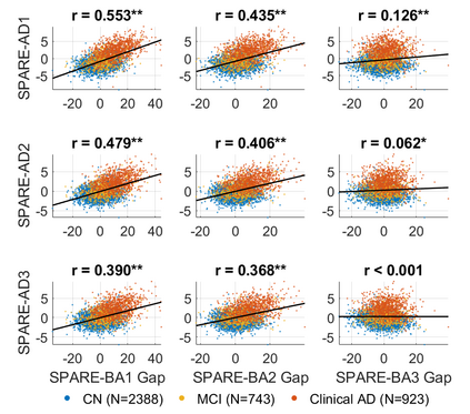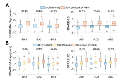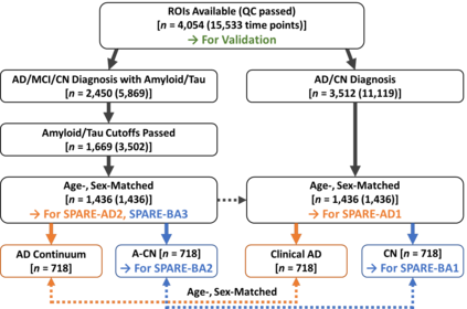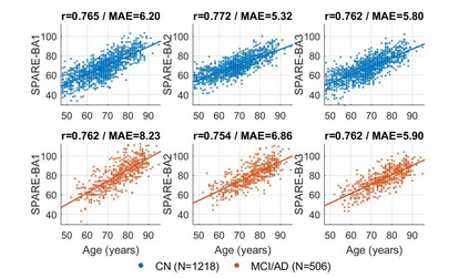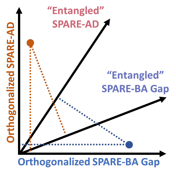
Neuroimaging biomarkers that distinguish between typical brain aging and Alzheimer's disease (AD) are valuable for determining how much each contributes to cognitive decline. Machine learning models can derive multi-variate brain change patterns related to the two processes, including the SPARE-AD (Spatial Patterns of Atrophy for Recognition of Alzheimer's Disease) and SPARE-BA (of Brain Aging) investigated herein. However, substantial overlap between brain regions affected in the two processes confounds measuring them independently. We present a methodology toward disentangling the two. T1-weighted MRI images of 4,054 participants (48-95 years) with AD, mild cognitive impairment (MCI), or cognitively normal (CN) diagnoses from the iSTAGING (Imaging-based coordinate SysTem for AGIng and NeurodeGenerative diseases) consortium were analyzed. First, a subset of AD patients and CN adults were selected based purely on clinical diagnoses to train SPARE-BA1 (regression of age using CN individuals) and SPARE-AD1 (classification of CN versus AD). Second, analogous groups were selected based on clinical and molecular markers to train SPARE-BA2 and SPARE-AD2: amyloid-positive (A+) AD continuum group (consisting of A+AD, A+MCI, and A+ and tau-positive CN individuals) and amyloid-negative (A-) CN group. Finally, the combined group of the AD continuum and A-/CN individuals was used to train SPARE-BA3, with the intention to estimate brain age regardless of AD-related brain changes. Disentangled SPARE models derived brain patterns that were more specific to the two types of the brain changes. Correlation between the SPARE-BA and SPARE-AD was significantly reduced. Correlation of disentangled SPARE-AD was non-inferior to the molecular measurements and to the number of APOE4 alleles, but was less to AD-related psychometric test scores, suggesting contribution of advanced brain aging to these scores.
翻译:将典型的大脑老化和阿尔茨海默氏病(AD)区分开来的生物标志中,将典型的大脑老化和阿尔茨海默氏病(AD)区分开来,对于确定每种疾病对认知下降的贡献有多大是有价值的。机器学习模型可以得出与这两个过程有关的多变大脑变化模式,包括SAPARE-AD(承认阿尔茨海默氏病的萎缩模式)和SPARE-BA(大脑进化)所有调查。然而,在两个过程中受影响的大脑区域之间有很大的重叠,我们展示了一种方法来分解这两种疾病。T1加权的MRI图像,4 054参与者(48-95岁)的T1加权MRI图像(48-95岁),有AD、轻度认知障碍(MCI)的大脑变化模式,或认知正常的 iSTAGA(以STem 和NAAAAAA(亚纳亚士亚士亚氏)的货币和SAAA(AAAAAAA)的亚运和AAAAAAAAAAA(进化)类和AAAAAAAAAAAAAAA(亚纳基)的肝和AAAA和AAAAAAAA(基)的肝和AAAAAAAA(基)的肝和AAA(基)的肝和AAAAAAAAAAA(基)的分级的分数的分数组,选择的分数组的肝和A和AAAAAAAAA)的分数(基)的分数组的分数)的分数组,选择的分。的分。和A,选择组和AA)的分的分数组的分。。
