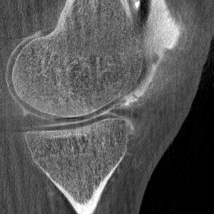
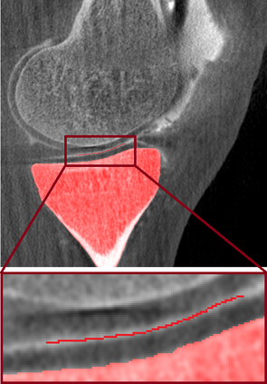
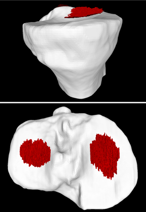
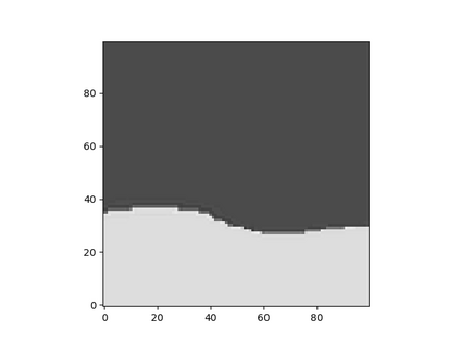
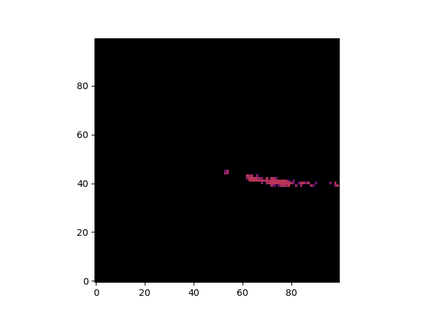
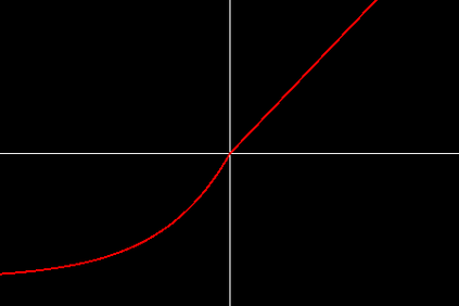
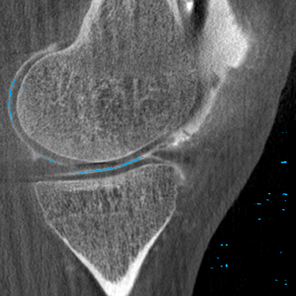
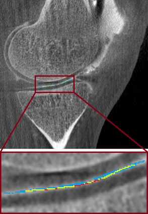
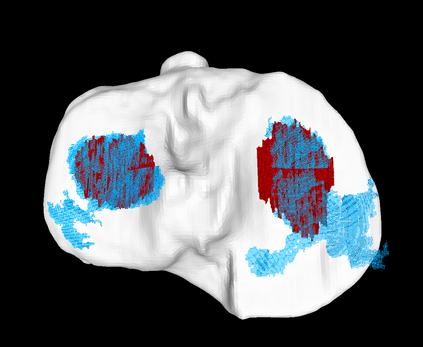
Analyzing knee cartilage thickness and strain under load can help to further the understanding of the effects of diseases like Osteoarthritis. A precise segmentation of the cartilage is a necessary prerequisite for this analysis. This segmentation task has mainly been addressed in Magnetic Resonance Imaging, and was rarely investigated on contrast-enhanced Computed Tomography, where contrast agent visualizes the border between femoral and tibial cartilage. To overcome the main drawback of manual segmentation, namely its high time investment, we propose to use a 3D Convolutional Neural Network for this task. The presented architecture consists of a V-Net with SeLu activation, and a Tversky loss function. Due to the high imbalance between very few cartilage pixels and many background pixels, a high false positive rate is to be expected. To reduce this rate, the two largest segmented point clouds are extracted using a connected component analysis, since they most likely represent the medial and lateral tibial cartilage surfaces. The resulting segmentations are compared to manual segmentations, and achieve on average a recall of 0.69, which confirms the feasibility of this approach.
翻译:分析膝盖软骨厚度和负荷下压力,有助于进一步了解骨质炎等疾病的影响。对软骨质炎的精确断裂是进行这一分析的一个必要先决条件。这一分离任务主要在磁共振成像中处理,很少通过对比强化的增强成像仪来调查,对比剂可以直观女性与湿软软骨质之间的边界。为了克服人工分割的主要缺陷,即高时投资,我们提议为此任务使用3D Convolual神经网络。所提出的结构包括一个带有SeLu激活功能的V-Net和Tversky损失功能。由于微小的软体像素和许多背景像素之间的高度不平衡,预计会出现高的假阳性率。为了降低这一比率,两个最大的断点云是通过一个连接的部件分析提取的,因为它们最有可能代表介质和较晚的土质板块表面。由此产生的断裂与人工分割方法相比,从而确定了平均的恢复0.69的可行性。

