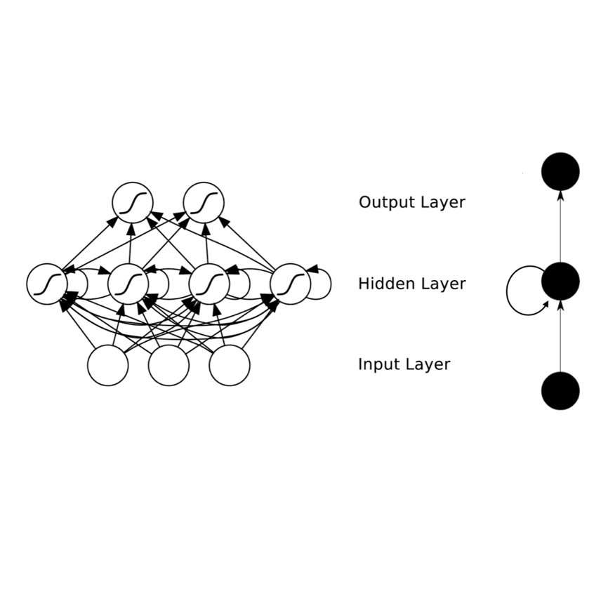
During the radiotherapy treatment of patients with lung cancer, the radiation delivered to healthy tissue around the tumor needs to be minimized, which is difficult because of respiratory motion and the latency of linear accelerator systems. In the proposed study, we first use the Lucas-Kanade pyramidal optical flow algorithm to perform deformable image registration of chest computed tomography scan images of four patients with lung cancer. We then track three internal points close to the lung tumor based on the previously computed deformation field and predict their position with a recurrent neural network (RNN) trained using real-time recurrent learning (RTRL) and gradient clipping. The breathing data is quite regular, sampled at approximately 2.5Hz, and includes artificial drift in the spine direction. The amplitude of the motion of the tracked points ranged from 12.0mm to 22.7mm. Finally, we propose a simple method for recovering and predicting 3D tumor images from the tracked points and the initial tumor image based on a linear correspondence model and Nadaraya-Watson non-linear regression. The root-mean-square error, maximum error, and jitter corresponding to the RNN prediction on the test set were smaller than the same performance measures obtained with linear prediction and least mean squares (LMS). In particular, the maximum prediction error associated with the RNN, equal to 1.51mm, is respectively 16.1% and 5.0% lower than the maximum error associated with linear prediction and LMS. The average prediction time per time step with RTRL is equal to 119ms, which is less than the 400ms marker position sampling time. The tumor position in the predicted images appears visually correct, which is confirmed by the high mean cross-correlation between the original and predicted images, equal to 0.955.
翻译:在对肺癌病人进行放射治疗期间,需要尽量减少肿瘤周围健康组织所受的辐射,这是由于呼吸运动和线性加速器系统的悬浮而很难做到的。在拟议的研究中,我们首先使用卢卡斯-卡纳德金字塔光学流算法对肺癌病人的4名病人进行胸腔可变图像登记,计算透视扫描图像,然后根据先前计算的畸形场,追踪接近肺癌病人的3个内部点,并用一个经常神经网络(RNNNN)预测他们的位置,这些网络经过实时经常性学习(RTRL)和梯度剪断的训练。呼吸数据非常经常,在大约2.5Hz取样,并包括脊椎方向的人工漂浮动。跟踪点的振动感光度从12.0毫米到22.7毫米不等。最后,我们提出了一个简单的方法,从跟踪点和最初肿瘤图中恢复并预测3D肿瘤图像,根据线性通信模型和Nadaraya-Watson非线性回归模型预测。根-正方位位置的定位是正常位置,在大约2.55H值位置上测得的测算,最大误差,最高误差和直线性测为最低测距值的测距值为最低测测距值为最低的测测距值,而测距值为最低测测测距值为最低测测距值为最低测测距值为最低。


