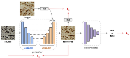
Stain color variation in histological images, caused by a variety of factors, is a challenge not only for the visual diagnosis of pathologists but also for cell segmentation algorithms. To eliminate the color variation, many stain normalization approaches have been proposed. However, most were designed for hematoxylin and eosin staining images and performed poorly on immunohistochemical staining images. Current cell segmentation methods systematically apply stain normalization as a preprocessing step, but the impact brought by color variation has not been quantitatively investigated yet. In this paper, we produced five groups of NeuN staining images with different colors. We applied a deep learning image-recoloring method to perform color transfer between histological image groups. Finally, we altered the color of a segmentation set and quantified the impact of color variation on cell segmentation. The results demonstrated the necessity of color normalization prior to subsequent analysis.
翻译:由多种因素造成的组织图象中的色素差异不仅对病理学家的视觉诊断是一个挑战,而且对细胞分解算法也是一个挑战。为了消除颜色差异,提出了许多污点正常化办法。然而,大多数是针对血氧素和叶素污渍图像设计的,对免疫生理化学污点图像的处理不力。目前细胞分解方法系统地将污点正常化作为处理前的一个步骤,但是对颜色差异带来的影响还没有进行定量调查。在本文中,我们制作了五组不同颜色的NeuN的染色图象。我们运用了一种深层学习的图像色化方法,在组织图象组之间进行色色转移。最后,我们改变了分解图案的颜色,并量化了颜色变化对细胞分解的影响。结果表明,在随后分析之前,必须实现颜色正常化。


