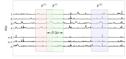
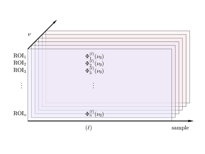
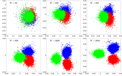
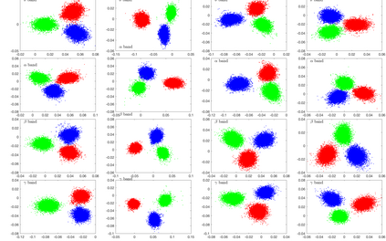
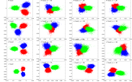
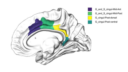
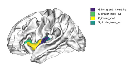
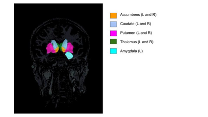
Meditation practices have been claimed to have a positive effect on the regulation of mood and emotion for quite some time by practitioners, and in recent times there has been a sustained effort to provide a more precise description of the changes induced by meditation on human brain. Longitudinal studies have reported morphological changes in cortical thickness and volume in selected brain regions due to meditation practice, which is interpreted as evidence for effectiveness of it beyond the subjective self reporting. Evidence based on real time monitoring of meditating brain by functional imaging modalities such as MEG or EEG remains a challenge. In this article we consider MEG data collected during meditation sessions of experienced Buddhist monks practicing focused attention (Samatha) and open monitoring (Vipassana) meditation, contrasted by resting state with eyes closed. The MEG data is first mapped to time series of brain activity averaged over brain regions corresponding to a standard Destrieux brain atlas, and further by bootstrapping and spectral analysis to data matrices representing a random sample of power spectral densities over bandwidths corresponding to $\alpha$, $\beta$, $\gamma$, and $\theta$ bands in the spectral range. We demonstrate using linear discriminant analysis (LDA) that the samples corresponding to different meditative or resting states contain enough fingerprints of the brain state to allow a separation between different states, and we identify the brain regions that appear to contribute to the separation. Our findings suggest that cingulate cortex, insular cortex and some of the internal structures, most notably accumbens, caudate and putamen nuclei, thalamus and amygdalae stand out as separating regions, which seems to correlate well with earlier findings based on longitudinal studies.
翻译:在一段时间里,实践者认为,模拟做法对调控情绪和情绪产生了积极的影响,最近,执业者认为对调控情绪和情绪产生了积极影响,而且最近还持续努力更准确地描述对人脑冥想引起的变化。纵向研究报告,由于冥想做法,某些大脑区域在骨骼厚度和体积方面的形态变化,这种变化被解释为其有效性的证据,超出了主观自我报告的范围。根据实时监测以功能成像模式(如MEG或EEG)对大脑进行冥想的证据仍然是一项挑战。在本篇文章中,我们认为,在有经验的佛教僧侣进行集中关注(萨玛塔)和开放监测(Vipassana)的冥想期间收集的MEG数据得到了更精确的描述。MEG数据首先被映射为大脑区域的平均大脑活动时间序列,相当于标准的Destrieux大脑图册,进一步通过靴子和光谱分析,显示我们内部和大脑结构内部结构的随机样本样本样本,显示我们内部和直径区域(我们之间的长期和直径区域)的直径分析似乎具有一定的频率。


