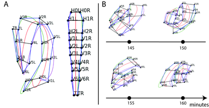
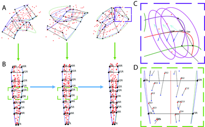
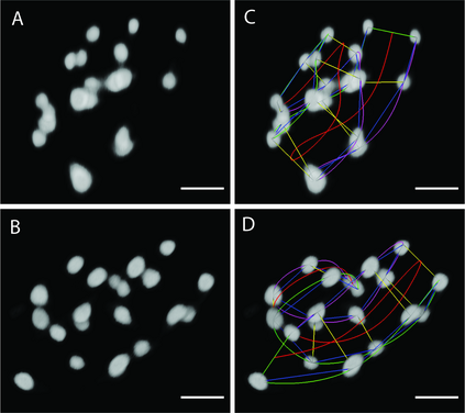
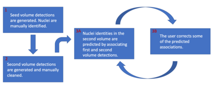
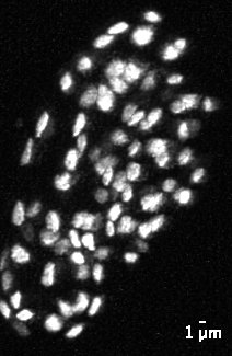
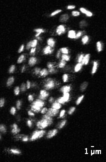
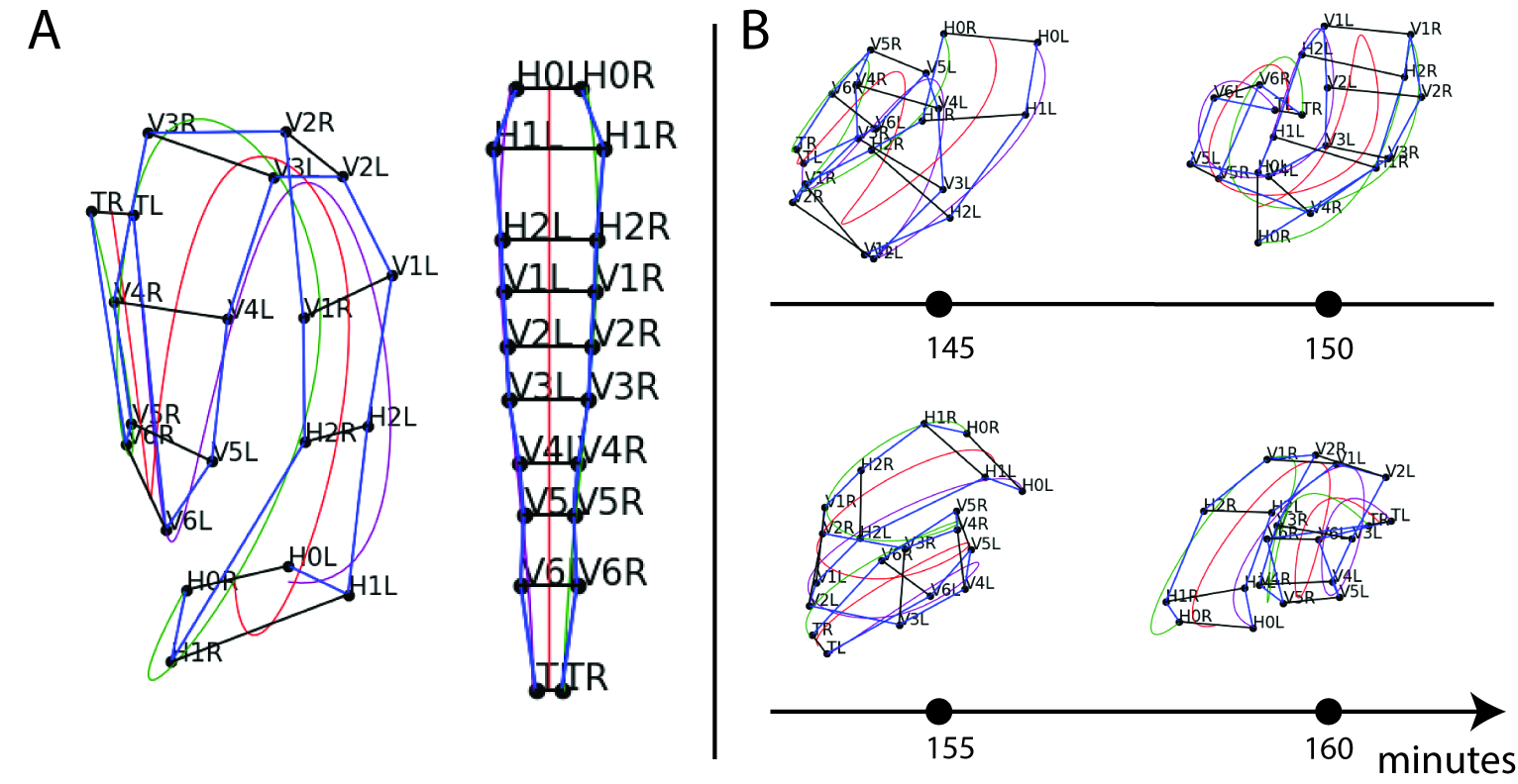
The nematode Caenorhabditis elegans (C. elegans) is used as a model organism to better understand developmental biology and neurobiology. C. elegans features an invariant cell lineage, which has been catalogued and observed using fluorescence microscopy images. However, established methods to track cells in late-stage development fail to generalize once sporadic muscular twitching has begun. We build upon methodology which uses skin cells as fiducial markers to carry out cell tracking despite random twitching. In particular, we present a cell nucleus segmentation and tracking procedure which was integrated into a 3D rendering GUI to improve efficiency in tracking cells across late-stage development. Results on images depicting aforementioned muscle cell nuclei across three test embryos suggest the fiducial markers in conjunction with a classic tracking paradigm overcome sporadic twitching.
翻译:C. 利根人具有一种无差异的细胞系系,使用荧光显微镜图像进行编目和观察。然而,在后期开发中跟踪细胞的既定方法未能在零星肌肉抽动开始后即普遍化。我们采用的方法是使用皮肤细胞作为纤维标志,进行细胞跟踪,尽管随机抽动。特别是,我们提出了一个细胞核分解和跟踪程序,纳入一个三维化图形界面,以提高跟踪细胞跨后阶段开发的效率。在三个测试胚胎上显示上述肌肉细胞核的图像的结果显示,在典型跟踪模式克服零星抽动的同时,还有典型的跟踪模式。

