

项目名称: 恶性肿瘤生物适形调强放疗PET/CT/MRI靶区智能勾画
项目编号: No.61271382
项目类型: 面上项目
立项/批准年度: 2013
项目学科: 无线电电子学、电信技术
项目作者: 刘国才
作者单位: 湖南大学
项目金额: 80万元
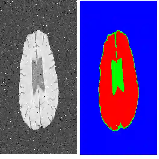
中文摘要: 在每年新增的两百多万癌症患者中,70%以上需要进行放射治疗。生物适形调强放疗成功实施的关键在生物靶区的高精度勾画,其本质是肿瘤分子生物特性功能影像PET图像分割,但肿瘤内部生物特性的各向异性特性和目前临床肿瘤PET图像固有的低空间解析度、强噪声与严重的部分容积效应,使高精度的肿瘤放疗生物靶区图像分割非常困难。因此,本项目提出联合肿瘤PET和高解析度的CT、MRI高阶纹理特征,采用各向异性向量值变分图像分割模型和水平集方法,集成临床专家肿瘤靶区勾画和组织病理知识,进行肿瘤靶区图像的高精度分割和智能勾画验证评估临床研究。为进一步提高靶区分割精度,计划将高解析度的肿瘤CT、MRI解剖结构信息集成到PET图像的最大后验概率统计重建和部分容积效应校正模型中,进行最大化PET图像空间解析度的统计重建优化方法和最小化肿瘤PET部分容积效应校正方法研究。对恶性肿瘤高精度放疗具有重要理论和临床应用研究价值。
中文关键词: 图像分割;PET/CT/MRI;放疗靶区勾画;放射治疗;头颈部肿瘤
英文摘要: Each year, the estimated number of new cancer cases in China is over 2 million and 70% of them need radiotherapy. In this era of high-precision biological conformal intensity-modulated radiotherapy, the accurate delieation of target volume with respect to tumor boundaries, shape, and volume is crucial, which may be achieved theoretically by the segmentation of PET tumor images. However, it is very difficult in practice due to the heterogeneity inside tumors and the low resolution , large noise and partial-volume effect(PVE) in PET tumor imaging. So, we propose that the PET, CT, and MRI textural characterizations such as coarseness, contrast, and busyness are extracted and used to the segmentation of PET tumor images via level set methodes and variational principles with clinical priors and a heterogeneous intensity model. The accuracy of the delineation of target volume is validated by pathologic examination. Moreover, the high-resolution image from CT or MRI is incorporated in the low-resolution PET image to yield a minimum PVE-corrected image and a maximum resolution PET reconstruction image by a maximum-a-posteriori approach.This project is very important to high-precision radiotherapy in theory and in clinical researches.
英文关键词: Image Segmentation;PET/CT/MRI;Delieation of target volume;Radiotherapy treatment planning;Head and neck cancer
成为VIP会员查看完整内容
相关内容
Arxiv
0+阅读 · 2022年4月20日
Arxiv
0+阅读 · 2022年4月20日
Arxiv
0+阅读 · 2022年4月15日
相关主题
相关VIP内容
相关资讯
相关论文
Arxiv
0+阅读 · 2022年4月20日
Arxiv
0+阅读 · 2022年4月20日
Arxiv
0+阅读 · 2022年4月15日